Model's Description:
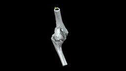
L. Elbow with Traumatic Fusion (VCU_3D_5443) 3D Model.
Download
L. Elbow with Traumatic Fusion (VCU_3D_5443) in various files formats such as Wavefront Object format, Autodesk FBX, DirectX 9.0, Stereo Lithography and HTML5 JSON format.
Left elbow (distal humerus and proximal ulna) with traumatic ankylosis from the American Civil War, collected by John Gouley. Specimen AFIP 1002831 from the National Museum of Health and Medicine (Silver Spring, MD). Model generated with the Segment Editor in 3D Slicer from a micro-computed tomography scan. 3D models can be downloaded from Morphosource at: https://www.morphosource.org/Detail/SpecimenDetail/Show/specimen_id/26332. Model generated by Terrie Simmons-Ehrhardt 3D Model is ready to download for free
, this model contains 1787840 polygons and 893522 vertices.
Model's rating is (3) based on 24 Votes.
Model's Description:
Humerus bone with landmarks and labels 3D Model.
Download Humerus bone with landmarks and labels in various files formats such as Wavefront Object format, Autodesk FBX, DirectX 9.0, Stereo Lithography and HTML5 JSON format.
Humerus bone showing landmarks including origins and insertions of muscles on it.
Click here if you want to see the model without labeling - Humerus bone with landmarks and labels - Buy Royalty Free 3D model by Anatomy by Doctor Jana (@docjana) 3D Model is ready to download for free, this model contains 3456 polygons.
Model's Description:
Brachial bone (humerus) cat 3D Model.
Download brachial bone (humerus) cat in various files formats such as Wavefront Object format, Autodesk FBX, DirectX 9.0, Stereo Lithography and HTML5 JSON format.
Right brachial bone (humerus) of a cat
size of the specimen: 93 x 7 x 18 mm
3D scanning performed with the structured light scanner “Artec Micro” - brachial bone (humerus) cat - 3D model by vetanatMunich 3D Model is ready to download for free, this model contains 50000 polygons.
Model's Description:
Humerus 3D Model.
Download Humerus in various files formats such as Wavefront Object format, Autodesk FBX, DirectX 9.0, Stereo Lithography and HTML5 JSON format.
3D scan of the right humerus
Captured with Einscan Pro
Captured and edited by: Madelyn Murphy
Copyright2019 BK Alsup & GM Fox - Humerus - Right, Labeled - 3D model by Bluelink Anatomy - University of Michigan (@bluelinkanatomy) 3D Model is ready to download for free, this model contains 201892 polygons.
Model's Description:
Lytic humeral lesion 3D Model.
Download Lytic humeral lesion in various files formats such as Wavefront Object format, Autodesk FBX, DirectX 9.0, Stereo Lithography and HTML5 JSON format.
-When a lytic lesion is suspected, the radiologist must keep in mind the possibility of a normal variant, such as a pseudocyst. Two common locations for pseudocysts are the humeral head and the calcaneus. The pseudocyst of the humeral head is typically located in the region of the greater tuberosity, while the pseudocyst of the calcaneus is typically located anteriorly.
-Proximal humerus chondrosarcoma is a rare localization of the common primary malignant cartilaginous tumor.
Reference:
1-https://appliedradiology.com/articles/general-approach-to-lytic-bone-lesions
2-https://www.researchgate.net/figure/Proximal-humerus-lytic-lesion-with-metaphyseal-and-epiphyseal-involvement_fig1_350678996 - Lytic humeral lesion - Buy Royalty Free 3D model by Ebers 3D Model is ready to download for free, this model contains 44318 polygons.
Model's Description:
Bones of The Upper Limb with Landmarks 3D Model.
Download Bones of The Upper Limb with Landmarks in various files formats such as Wavefront Object format, Autodesk FBX, DirectX 9.0, Stereo Lithography and HTML5 JSON format.
It is difficult to find a model of human bones which has proper anatomical landmarks online so I started the osteology project. I am creating detailed sculpts of human body and then optimizing them to run even on low end devices.
The goal is to create high quality assets with good subdividable topology. If you are searching for a good sculpted and textured models of human body which is both medically accurate and visually beautiful you have come to the right place!
All the models in the project will be pure quads with no tris and retopologized by hand to squeeze out the last vertex without losing the overall look of the model.
All the meshes are created from high resolution sculpts I made from years of study on the human anatomy. This is a single person endeavor so please be patient with me. Releases might be slow but I am working on them.
For any business enquiries or custom models contact me on doctorjana57@gmail.com 3D Model is ready to download for free, this model contains 48076 polygons.
Model's Description:
Tibia/Fibula with Osteomyelitis (VCU_3D_5442) 3D Model.
Download Tibia/Fibula with Osteomyelitis (VCU_3D_5442) in various files formats such as Wavefront Object format, Autodesk FBX, DirectX 9.0, Stereo Lithography and HTML5 JSON format.
Right distal tibia/fibula with osteomyelitis/healed fracture from the American Civil War; patient William Holmes Co. D 18th Massachusetts, injured at Fredericksburg, VA Dec 13, 1862. Specimen AFIP 1002694 from the National Museum of Health and Medicine (Silver Spring, MD).
Model generated with the Segment Editor in 3D Slicer from a micro-computed tomography scan. 3D models can be downloaded from Morphosource at: https://www.morphosource.org/Detail/SpecimenDetail/Show/specimen_id/31978.
Model generated by Terrie Simmons-Ehrhardt 3D Model is ready to download for free, this model contains 2828626 polygons.
Model's Description:
Surgery Light Cartoon Style 3D Model.
Download Surgery Light Cartoon Style in various files formats such as Wavefront Object format, Autodesk FBX, DirectX 9.0, Stereo Lithography and HTML5 JSON format.
Surgery light for pathology ward. Part of Red Awakening game (http://www.red-awakening.com) - Surgery Light Cartoon Style - 3D model by varnenche 3D Model is ready to download for free, this model contains 1463 polygons.
Model's Description:
Lytic Lesions of spine 3D Model.
Download Lytic Lesions of spine in various files formats such as Wavefront Object format, Autodesk FBX, DirectX 9.0, Stereo Lithography and HTML5 JSON format.
-the vertebra is divided anatomically into the vertebral body anteriorly and the neural arch posteriorly. The neural arch is composed of bilateral pedicles, pars interarticularis, and laminae; from the neural arch arise the spinous process, bilateral transverse processes, and bilateral superior and inferior articulating processes. These anatomic components are known collectively as the posterior elements of the vertebra.
-Myeloma bone disease can result not only in fractures, but in pressure on the spinal cord (spinal cord compression), the need for surgery to prevent or repair broken bones, and/or the need for radiation treatments to the bone to control the myeloma and relieve pain. These are collectively known as “skeletal-related events.”
Reference:-
1-https://journals.lww.com/cdronline/Abstract/2017/11150/Lytic_Lesions_of_the_Posterior_Elements_of_the.1.aspx
2-https://www.myeloma.org/bone-disease - Lytic Lesions of spine - Buy Royalty Free 3D model by Ebers 3D Model is ready to download for free, this model contains 56920 polygons.
Model's Description:
Lytic Lesions of Rib 3D Model.
Download Lytic Lesions of Rib in various files formats such as Wavefront Object format, Autodesk FBX, DirectX 9.0, Stereo Lithography and HTML5 JSON format.
-In skeletal tumors, metastatic bone tumors have the most common occurrence [1] and primary bone tumors of chest wall accounts for only 5 - 8 % of all bony tumors.
-Of these approximately 95% occur in ribs and remainder in the sternum.
Reference :-
https://www.fortunejournals.com/articles/expansile-lytic-lesions-of-rib-two-rare-case-reports.html - Lytic Lesions of Rib - Buy Royalty Free 3D model by Ebers 3D Model is ready to download for free, this model contains 118150 polygons.
Model's Description:
Left Frontosphenoidal Craniosynostosis 3D Model.
Download Left Frontosphenoidal Craniosynostosis in various files formats such as Wavefront Object format, Autodesk FBX, DirectX 9.0, Stereo Lithography and HTML5 JSON format.
An infant cranium and mandible (6 months old) with left frontosphenoidal craniosynostosis. Small gaps between cranial bones were filled to make it easier to 3D print. The mandible was also solidified to make 3D printing easier (unerupted teeth are not visible)–central mandibular incisors are erupted. You can also cut the cranium before 3D printing to have access to the endocranial anatomy.
I segmented this from a computed tomography scan from Embodi3D.
Download includes beige PLY of the cranium and mandible combined. The additional file contains the separated STL models for 3D printing 3D Model is ready to download for free, this model contains 339948 polygons.
Model's Description:
Taylor Mtn, Idaho Mule Deer Skull 3D Model.
Download Taylor Mtn in various files formats such as Wavefront Object format, Autodesk FBX, DirectX 9.0, Stereo Lithography and HTML5 JSON format.
Taylor Mtn, Idaho Mule Deer Skull - 3D model by Idaho Virtualization Laboratory (@ivlpaleontology) 3D Model is ready to download for free, this model contains 320916 polygons.
Model's Description:
Human spinal column. 3D Model.
Download Human spinal column. in various files formats such as Wavefront Object format, Autodesk FBX, DirectX 9.0, Stereo Lithography and HTML5 JSON format.
Segmented complete human vertebral column, from a CT scan on the National Cancer Imaging Archive (ID# 0522x0132).
This was segmented manually using the freeware program Stradview.
The cervical, thoracic and lumbar vertebrae are coloured differently to aid in identification 3D Model is ready to download for free, this model contains 206904 polygons.
Model's Description:
Sumatran Tiger Skeleton 3D Model.
Download Sumatran Tiger Skeleton in various files formats such as Wavefront Object format, Autodesk FBX, DirectX 9.0, Stereo Lithography and HTML5 JSON format.
The Sumatran Tiger subspecies is critically endangered with as few as 400 living in the island of Sumatra, Indonesia.
3d modeling based on the University of Georgia College video on youtube and various drawings and photos 3D Model is ready to download for free, this model contains 100718 polygons.
Model's Description:
Occipital 3D Model.
Download Occipital in various files formats such as Wavefront Object format, Autodesk FBX, DirectX 9.0, Stereo Lithography and HTML5 JSON format.
Human occipital from the Osteology Collection, Centre for Anatomy and Human Identification (CAHID), University of Dundee.
Created by: Staff
Hardware: Artec Space Spider
Software: Artec Studio 15
Models are all produced for teaching. We have a collection of models only visible to our students please contact us for more information. https://www.dundee.ac.uk/cahid - Occipital - 3D model by University of Dundee, CAHID (@anatomy_dundee) 3D Model is ready to download for free, this model contains 849458 polygons.
Model's Description:
Skeleton 3D Model.
Download Skeleton in various files formats such as Wavefront Object format, Autodesk FBX, DirectX 9.0, Stereo Lithography and HTML5 JSON format.
This is a basic male skeleton. I modelled it in order to use it for anatomy stydies. Mi intencion is to rig is later on and be able to pose it. The mesh comes in a single object but its all separated pieces. If you want to rig it and pose it you’ll have to separate de pieces and organice them. It has no UVs or textures. If you use it you may find mistakes or things to improve. I would apreciate very much your feed back.
You can see more about this model on:
https://www.artstation.com/artwork/LkmLk - Skeleton - Download Free 3D model by Diego Luján García (@diegoluga) 3D Model is ready to download for free, this model contains 378572 polygons.
Model's Description:
Ulna (ulna) cat 3D Model.
Download ulna (ulna) cat in various files formats such as Wavefront Object format, Autodesk FBX, DirectX 9.0, Stereo Lithography and HTML5 JSON format.
Right ulna (ulna) of a cat
size of the specimen: 91 x 9 x 7 mm
3D scanning performed with the structured light scanner “Artec Space Spider” - ulna (ulna) cat - 3D model by vetanatMunich 3D Model is ready to download for free, this model contains 43288 polygons.
Model's Description:
Human Forearm 3D Model.
Download Human Forearm in various files formats such as Wavefront Object format, Autodesk FBX, DirectX 9.0, Stereo Lithography and HTML5 JSON format.
3D scan of human skeleton replica. Right side, forearm/ lower arm bones - ulna and radius.
“The forearm is the region of the upper limb between the elbow and the wrist. The term forearm is used in anatomy to distinguish it from the arm, a word which is most often used to describe the entire appendage of the upper limb, but which in anatomy, technically, means only the region of the upper arm, whereas the lower “arm” is called the forearm. It is homologous to the region of the leg that lies between the knee and the ankle joints, the crus. The forearm contains two long bones, the radius and the ulna, forming the radioulnar joint. The interosseous membrane connects these bones. Ultimately, the forearm is covered by skin, the anterior surface usually being less hairy than the posterior surface.” - Wikipedia
Captured by K. Jarvinen with Artec Spider 3D Scanner https://www.artec3d.com/portable-3d-scanners/artec-spider-v2 - Human Forearm - ulna and radius (replica) - 3D model by RISD Nature Lab (@RISDNaturelab) 3D Model is ready to download for free, this model contains 1000000 polygons.
Model's Description:
Pronation/supination 3D Model.
Download Pronation/supination in various files formats such as Wavefront Object format, Autodesk FBX, DirectX 9.0, Stereo Lithography and HTML5 JSON format.
This is an animation of the arm bones: humerus, ulna and radius showing how the elbow joint works during pronation/supination with along with flexion and extension.
I made it as part of a series of blog posts I’m working on to describe anatomy for artists which you can find here:
https://pearsetoomey.com/bones-of-the-arm/ - Pronation/supination - arm bones animated - 3D model by pearsetoomey 3D Model is ready to download for free, this model contains 17822 polygons.
Model's Description:
Horse bone 3D Model.
Download Horse bone in various files formats such as Wavefront Object format, Autodesk FBX, DirectX 9.0, Stereo Lithography and HTML5 JSON format.
Bone of an extinct horse breed from southern Germany. Original scan counts about 5.000.000 polygons. For more information visit http://lucabrunke.de
Cycles render of the high poly bone 3D Model is ready to download for free, this model contains 6090 polygons.
Model's Description:
Creation Of Adam 3D Model.
Download Creation Of Adam in various files formats such as Wavefront Object format, Autodesk FBX, DirectX 9.0, Stereo Lithography and HTML5 JSON format.
I thought I’d recreate that famous frame of Micheal Aaneglo’s creation of Adam in 3D, It was supposed to be a quick anatomy study, But I’m glad I ended up texturing it, check The full project on my artstation and let me know what you think 3D Model is ready to download for free, this model contains 759392 polygons.
Model's Description:
Monster-hand 3D Model.
Download Monster-hand in various files formats such as Wavefront Object format, Autodesk FBX, DirectX 9.0, Stereo Lithography and HTML5 JSON format.
Monster-hand
Game assets
3D MODEL BY https://www.facebook.com/jm.cigarza
JM CIgarza - Monster-hand - 3D model by NORBERTO-3D (@norberto3d) 3D Model is ready to download for free, this model contains 144774 polygons.
Model's Description:
Low poly hand painted anatomy model 3D Model.
Download Low poly hand painted anatomy model in various files formats such as Wavefront Object format, Autodesk FBX, DirectX 9.0, Stereo Lithography and HTML5 JSON format.
Before buying watch video demo:
https://youtu.be/nQd2Z6uispM
Low poly hand painted human Anatomy Model. Include all major anatomical parts. All the muscles and tendons, Skeleton, Organs and glands, Circulatory system, Nervous system, Lymphatic system, respiratory system etc.
Some object have only single side (like some muscles, veins etc.) to reduce poly count.
Textures are handpainted. Light is painted in to texture so model work best with flat lighting (this also improve speed)
I am not medically trained so some smaller parts may be inconsistent as different sources describe them differently.
Model poly count:
Verts: 260 104
Faces: 201 771
Tris: 368 069
All textures are 4k .png
File format: .blend .fbx .obj
Blend fille with al gruped and named parts is in .zip archive - Low poly hand painted anatomy model - Buy Royalty Free 3D model by graft 3D Model is ready to download for free, this model contains 362961 polygons.
 3D Model")


 cat")


")


 cat")


