Model's Description:
Fetal eye cross section week sixteen 3D Model, Fully textured with UV Mapping and materials.
Download
Fetal eye cross section week sixteen in various files formats such as Wavefront Object format, Autodesk FBX, DirectX 9.0, Stereo Lithography and HTML5 JSON format.
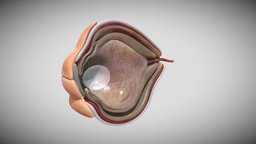
Although eye development can be considered from an embryological perspective to commence around day 22, when the optic sulci (optic primordium) appear as shallow grooves or pits in the inner aspect of the neural plate or neural folds ( Fig. 2-1A ) and the embryo is around 2 mm in length with eight somites, the group of cells that constitute the eye primordium or eye field have already begun to express a set of ‘eye field transcription factors’ (EFTFs) that are highly conserved in our evolutionary ancestory References :- 1- Larsen's Human Embryology textbook ( Sixth Edition ). 2- the anatomy of human embryo electron microscope textbook ( 1st Edition ) . 3-langman medical embryology textbook( Fourteenth Edition ) 3D Model is ready to download for free
with 51 Texture files attached, this model contains 110180 polygons and 55108 vertices.
Model's rating is (3) based on 51 Votes.
Download : (1,016 Hits)
for OBJ, FBX, GLTF, GLB, DAE and other formats... or use
Model's Description:
Osteolytic lesion 3D Model, Optimized by RigModels.com, Fully textured with UV Mapping and materials.
Download Osteolytic lesion in various files formats such as Wavefront Object format, Autodesk FBX, DirectX 9.0, Stereo Lithography and HTML5 JSON format.
Bone tumors are mostly benign. The most important determinants in imaging of bone tumors are morphology on plain radiograph (well-defined lytic, ill-defined lytic, and sclerotic lesions) and age of the patient at presentation.
-Well-defined osteolytic bone tumors and tumor-like lesions have a plethora of differentials in different age groups. For simplicity, a widely used mnemonic for lytic bone lesions is extremely helpful: FEGNOMASHIC. We have attempted to describe the most characteristic features of each of these tumors.
Reference:-
https://pubmed.ncbi.nlm.nih.gov/30969659/ - Osteolytic lesion - Buy Royalty Free 3D model by Ebers 3D Model is ready to download for free, this model contains 53070 polygons.
Model's Description:
Lytic humeral lesion 3D Model, Optimized by RigModels.com, Fully textured with UV Mapping and materials.
Download Lytic humeral lesion in various files formats such as Wavefront Object format, Autodesk FBX, DirectX 9.0, Stereo Lithography and HTML5 JSON format.
-When a lytic lesion is suspected, the radiologist must keep in mind the possibility of a normal variant, such as a pseudocyst. Two common locations for pseudocysts are the humeral head and the calcaneus. The pseudocyst of the humeral head is typically located in the region of the greater tuberosity, while the pseudocyst of the calcaneus is typically located anteriorly.
-Proximal humerus chondrosarcoma is a rare localization of the common primary malignant cartilaginous tumor.
Reference:
1-https://appliedradiology.com/articles/general-approach-to-lytic-bone-lesions
2-https://www.researchgate.net/figure/Proximal-humerus-lytic-lesion-with-metaphyseal-and-epiphyseal-involvement_fig1_350678996 - Lytic humeral lesion - Buy Royalty Free 3D model by Ebers 3D Model is ready to download for free, this model contains 44318 polygons.
Model's Description:
Chromosome Structure 3D Model, Optimized by RigModels.com, Fully textured with UV Mapping and materials.
Download Chromosome Structure in various files formats such as Wavefront Object format, Autodesk FBX, DirectX 9.0, Stereo Lithography and HTML5 JSON format.
A chromosome is a long DNA molecule with part or all of the genetic material of an organism. Most eukaryotic chromosomes include packaging proteins called histones which, aided by chaperone proteins, bind to and condense the DNA molecule to maintain its integrity. These chromosomes display a complex three-dimensional structure, which plays a significant role in transcriptional regulation 3D Model is ready to download for free, this model contains 272640 polygons.
Model's Description:
Earth Globe 3D Model, Optimized by RigModels.com, Fully textured with UV Mapping and materials.
Download Earth Globe in various files formats such as Wavefront Object format, Autodesk FBX, DirectX 9.0, Stereo Lithography and HTML5 JSON format.
Earth Globe 3D Model is ready to download for free, this model contains 9692 polygons.
Model's Description:
Orofacial anatomy with blood and nerve supply 3D Model, Optimized by RigModels.com, Fully textured with UV Mapping and materials.
Download Orofacial anatomy with blood and nerve supply in various files formats such as Wavefront Object format, Autodesk FBX, DirectX 9.0, Stereo Lithography and HTML5 JSON format.
Visualization of the orofacial anatomy showing the blood and nerve supply of this area.
Kindly check our channel for more models on the following link www.sketchfab.com/ebers - Orofacial anatomy with blood and nerve supply - Buy Royalty Free 3D model by Ebers 3D Model is ready to download for free, this model contains 758582 polygons.
Model's Description:
Benign prostatic hyperplasia 3D Model, Optimized by RigModels.com, Fully textured with UV Mapping and materials.
Download Benign prostatic hyperplasia in various files formats such as Wavefront Object format, Autodesk FBX, DirectX 9.0, Stereo Lithography and HTML5 JSON format.
Benign prostatic hyperplasia is a condition in which the prostate gland grows larger. The enlarged prostate may block or slow the passage of urine from the urethra 3D Model is ready to download for free, this model contains 44848 polygons.
Model's Description:
Male Reproductive System Week Sixteen 3D Model, Optimized by RigModels.com, Fully textured with UV Mapping and materials.
Download Male Reproductive System Week Sixteen in various files formats such as Wavefront Object format, Autodesk FBX, DirectX 9.0, Stereo Lithography and HTML5 JSON format.
In the first stage of gonadal development, it is impossible to distinguish between the male and female gonad. Thus, it is known as the indifferent stage.
The gonads begin as genital ridges – a pair of longitudinal ridges derived from intermediate mesoderm and overlying epithelium. They initially do not contain any germ cells.
Male and female reproductive systems develop in close relation to the urinary tract. Until approximately 7 weeks gestation, the human embryo remains ually bipotential. Subsequently, in males, testis-inducing factors cause differentiation from the default female phenotype.
References :
1- Netters atals human emberyology (Updated Edition).
2-Human embryology and development biology (6th Edition) .
3- the anatomy of human embryo electron microscope ( 1st Edition ) 3D Model is ready to download for free, this model contains 40160 polygons.
Model's Description:
Fetal ear cross-section week sixteen 3D Model, Optimized by RigModels.com, Fully textured with UV Mapping and materials.
Download Fetal ear cross-section week sixteen in various files formats such as Wavefront Object format, Autodesk FBX, DirectX 9.0, Stereo Lithography and HTML5 JSON format.
The ear is a composite structure with multiple embryonic origins , the external and middle ears arise from the first and second pharyngeal arches and the intervening pharyngeal cleft , membrane and pouch.
Reference :
1-Netter atlas human embryology (Updated Edition)
2-the anatomy of human embryo electron microscope textbook ( 1st Edition ) 3D Model is ready to download for free, this model contains 49826 polygons.