Model's Description:
L. Elbow with Traumatic Fusion (VCU_3D_5443) 3D Model.
Download
L. Elbow with Traumatic Fusion (VCU_3D_5443) in various files formats such as Wavefront Object format, Autodesk FBX, DirectX 9.0, Stereo Lithography and HTML5 JSON format.
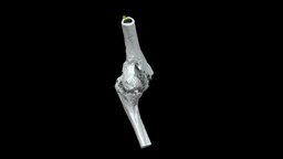
Left elbow (distal humerus and proximal ulna) with traumatic ankylosis from the American Civil War, collected by John Gouley. Specimen AFIP 1002831 from the National Museum of Health and Medicine (Silver Spring, MD). Model generated with the Segment Editor in 3D Slicer from a micro-computed tomography scan. 3D models can be downloaded from Morphosource at: https://www.morphosource.org/Detail/SpecimenDetail/Show/specimen_id/26332. Model generated by Terrie Simmons-Ehrhardt 3D Model is ready to download for free
, this model contains 1787840 polygons and 893522 vertices.
Model's rating is (3) based on 39 Votes.
Model's Description:
Humerus 3D Model.
Download Humerus in various files formats such as Wavefront Object format, Autodesk FBX, DirectX 9.0, Stereo Lithography and HTML5 JSON format.
3D scan of the right humerus
Captured with Einscan Pro
Captured and edited by: Madelyn Murphy
Copyright2019 BK Alsup & GM Fox - Humerus - Right, Labeled - 3D model by Bluelink Anatomy - University of Michigan (@bluelinkanatomy) 3D Model is ready to download for free, this model contains 201892 polygons.
Model's Description:
Humerus bone with landmarks and labels 3D Model.
Download Humerus bone with landmarks and labels in various files formats such as Wavefront Object format, Autodesk FBX, DirectX 9.0, Stereo Lithography and HTML5 JSON format.
Humerus bone showing landmarks including origins and insertions of muscles on it.
Click here if you want to see the model without labeling - Humerus bone with landmarks and labels - Buy Royalty Free 3D model by Anatomy by Doctor Jana (@docjana) 3D Model is ready to download for free, this model contains 3456 polygons.
Model's Description:
Brachial bone (humerus) cat 3D Model.
Download brachial bone (humerus) cat in various files formats such as Wavefront Object format, Autodesk FBX, DirectX 9.0, Stereo Lithography and HTML5 JSON format.
Right brachial bone (humerus) of a cat
size of the specimen: 93 x 7 x 18 mm
3D scanning performed with the structured light scanner “Artec Micro” - brachial bone (humerus) cat - 3D model by vetanatMunich 3D Model is ready to download for free, this model contains 50000 polygons.
Model's Description:
Bones of The Upper Limb with Landmarks 3D Model.
Download Bones of The Upper Limb with Landmarks in various files formats such as Wavefront Object format, Autodesk FBX, DirectX 9.0, Stereo Lithography and HTML5 JSON format.
It is difficult to find a model of human bones which has proper anatomical landmarks online so I started the osteology project. I am creating detailed sculpts of human body and then optimizing them to run even on low end devices.
The goal is to create high quality assets with good subdividable topology. If you are searching for a good sculpted and textured models of human body which is both medically accurate and visually beautiful you have come to the right place!
All the models in the project will be pure quads with no tris and retopologized by hand to squeeze out the last vertex without losing the overall look of the model.
All the meshes are created from high resolution sculpts I made from years of study on the human anatomy. This is a single person endeavor so please be patient with me. Releases might be slow but I am working on them.
For any business enquiries or custom models contact me on doctorjana57@gmail.com 3D Model is ready to download for free, this model contains 48076 polygons.
Model's Description:
Lytic humeral lesion 3D Model.
Download Lytic humeral lesion in various files formats such as Wavefront Object format, Autodesk FBX, DirectX 9.0, Stereo Lithography and HTML5 JSON format.
-When a lytic lesion is suspected, the radiologist must keep in mind the possibility of a normal variant, such as a pseudocyst. Two common locations for pseudocysts are the humeral head and the calcaneus. The pseudocyst of the humeral head is typically located in the region of the greater tuberosity, while the pseudocyst of the calcaneus is typically located anteriorly.
-Proximal humerus chondrosarcoma is a rare localization of the common primary malignant cartilaginous tumor.
Reference:
1-https://appliedradiology.com/articles/general-approach-to-lytic-bone-lesions
2-https://www.researchgate.net/figure/Proximal-humerus-lytic-lesion-with-metaphyseal-and-epiphyseal-involvement_fig1_350678996 - Lytic humeral lesion - Buy Royalty Free 3D model by Ebers 3D Model is ready to download for free, this model contains 44318 polygons.
Model's Description:
Tibia/Fibula with Osteomyelitis (VCU_3D_5442) 3D Model.
Download Tibia/Fibula with Osteomyelitis (VCU_3D_5442) in various files formats such as Wavefront Object format, Autodesk FBX, DirectX 9.0, Stereo Lithography and HTML5 JSON format.
Right distal tibia/fibula with osteomyelitis/healed fracture from the American Civil War; patient William Holmes Co. D 18th Massachusetts, injured at Fredericksburg, VA Dec 13, 1862. Specimen AFIP 1002694 from the National Museum of Health and Medicine (Silver Spring, MD).
Model generated with the Segment Editor in 3D Slicer from a micro-computed tomography scan. 3D models can be downloaded from Morphosource at: https://www.morphosource.org/Detail/SpecimenDetail/Show/specimen_id/31978.
Model generated by Terrie Simmons-Ehrhardt 3D Model is ready to download for free, this model contains 2828626 polygons.
Model's Description:
Surgery Light Cartoon Style 3D Model.
Download Surgery Light Cartoon Style in various files formats such as Wavefront Object format, Autodesk FBX, DirectX 9.0, Stereo Lithography and HTML5 JSON format.
Surgery light for pathology ward. Part of Red Awakening game (http://www.red-awakening.com) - Surgery Light Cartoon Style - 3D model by varnenche 3D Model is ready to download for free, this model contains 1463 polygons.
Model's Description:
Left Frontosphenoidal Craniosynostosis 3D Model.
Download Left Frontosphenoidal Craniosynostosis in various files formats such as Wavefront Object format, Autodesk FBX, DirectX 9.0, Stereo Lithography and HTML5 JSON format.
An infant cranium and mandible (6 months old) with left frontosphenoidal craniosynostosis. Small gaps between cranial bones were filled to make it easier to 3D print. The mandible was also solidified to make 3D printing easier (unerupted teeth are not visible)–central mandibular incisors are erupted. You can also cut the cranium before 3D printing to have access to the endocranial anatomy.
I segmented this from a computed tomography scan from Embodi3D.
Download includes beige PLY of the cranium and mandible combined. The additional file contains the separated STL models for 3D printing 3D Model is ready to download for free, this model contains 339948 polygons.
 3D Model")


 cat")


")


