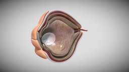
Model's Description:
Fetal eye cross section week sixteen 3D Model, Fully textured with UV Mapping and materials.
Download
Fetal eye cross section week sixteen in various files formats such as Wavefront Object format, Autodesk FBX, DirectX 9.0, Stereo Lithography and HTML5 JSON format.
Although eye development can be considered from an embryological perspective to commence around day 22, when the optic sulci (optic primordium) appear as shallow grooves or pits in the inner aspect of the neural plate or neural folds ( Fig. 2-1A ) and the embryo is around 2 mm in length with eight somites, the group of cells that constitute the eye primordium or eye field have already begun to express a set of ‘eye field transcription factors’ (EFTFs) that are highly conserved in our evolutionary ancestory References :- 1- Larsen's Human Embryology textbook ( Sixth Edition ). 2- the anatomy of human embryo electron microscope textbook ( 1st Edition ) . 3-langman medical embryology textbook( Fourteenth Edition ) 3D Model is ready to download for free
with 51 Texture files attached, this model contains 110180 polygons and 55108 vertices.
Model's rating is (3) based on 30 Votes.
Download : (1,028 Hits)
for OBJ, FBX, GLTF, GLB, DAE and other formats... or use
Model's Description:
Liver,Spleen and Pancreas Week Eight 3D Model, Optimized by RigModels.com, Fully textured with UV Mapping and materials.
Download Liver in various files formats such as Wavefront Object format, Autodesk FBX, DirectX 9.0, Stereo Lithography and HTML5 JSON format.
The epithelium of and the parenchyma of glands associated with the digestive tract (e.g., liver and pancreas) are derived from endoderm. The muscular walls of the digestive tract (lamina propria, muscularis mucosae, submucosa, muscularis externa, adventitia and/or serosa) are derived from splanchnic mesoderm.
Reference
Larsen’s Human Embryology textbook ( Sixth Edition ) 3D Model is ready to download for free, this model contains 7724 polygons.
Model's Description:
Lung Cancer 3D Model, Optimized by RigModels.com, Fully textured with UV Mapping and materials.
Download Lung Cancer in various files formats such as Wavefront Object format, Autodesk FBX, DirectX 9.0, Stereo Lithography and HTML5 JSON format.
Lung cancer or bronchogenic carcinoma refers to tumors originating in the lung parenchyma or within the bronchi. It is one of the leading causes of cancer-related deaths in the United States. Since 1987, lung cancer has been responsible for more deaths in women than breast cancer. It is estimated that there are 225,000 new cases of lung cancer in the United States annually, and approximately 160,000 die because of lung cancer. It is interesting to note that lung cancer was a relatively rare disease at the beginning of the 20th century. Its dramatic rise in later decades is attributable primarily to the increase in smoking among both males and females.
Reference
Siddiqui F, Vaqar S, Siddiqui AH. Lung Cancer. [Updated 2022 Dec 5]. In: StatPearls [Internet]. Treasure Island (FL): StatPearls Publishing; 2022 Jan-. Available from: https://www.ncbi.nlm.nih.gov/books/NBK482357/ - Lung Cancer - Buy Royalty Free 3D model by Ebers 3D Model is ready to download for free, this model contains 595154 polygons.