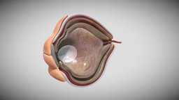
Model's Description:
Fetal eye cross section week sixteen 3D Model, Fully textured with UV Mapping and materials.
Download
Fetal eye cross section week sixteen in various files formats such as Wavefront Object format, Autodesk FBX, DirectX 9.0, Stereo Lithography and HTML5 JSON format.
Although eye development can be considered from an embryological perspective to commence around day 22, when the optic sulci (optic primordium) appear as shallow grooves or pits in the inner aspect of the neural plate or neural folds ( Fig. 2-1A ) and the embryo is around 2 mm in length with eight somites, the group of cells that constitute the eye primordium or eye field have already begun to express a set of ‘eye field transcription factors’ (EFTFs) that are highly conserved in our evolutionary ancestory References :- 1- Larsen's Human Embryology textbook ( Sixth Edition ). 2- the anatomy of human embryo electron microscope textbook ( 1st Edition ) . 3-langman medical embryology textbook( Fourteenth Edition ) 3D Model is ready to download for free
with 51 Texture files attached, this model contains 110180 polygons and 55108 vertices.
Model's rating is (3) based on 32 Votes.
Model's Description:
Lytic humeral lesion 3D Model.
Download Lytic humeral lesion in various files formats such as Wavefront Object format, Autodesk FBX, DirectX 9.0, Stereo Lithography and HTML5 JSON format.
-When a lytic lesion is suspected, the radiologist must keep in mind the possibility of a normal variant, such as a pseudocyst. Two common locations for pseudocysts are the humeral head and the calcaneus. The pseudocyst of the humeral head is typically located in the region of the greater tuberosity, while the pseudocyst of the calcaneus is typically located anteriorly.
-Proximal humerus chondrosarcoma is a rare localization of the common primary malignant cartilaginous tumor.
Reference:
1-https://appliedradiology.com/articles/general-approach-to-lytic-bone-lesions
2-https://www.researchgate.net/figure/Proximal-humerus-lytic-lesion-with-metaphyseal-and-epiphyseal-involvement_fig1_350678996 - Lytic humeral lesion - Buy Royalty Free 3D model by Ebers 3D Model is ready to download for free, this model contains 44318 polygons.
Model's Description:
Liver,Spleen and Pancreas Week Eight 3D Model.
Download Liver in various files formats such as Wavefront Object format, Autodesk FBX, DirectX 9.0, Stereo Lithography and HTML5 JSON format.
The epithelium of and the parenchyma of glands associated with the digestive tract (e.g., liver and pancreas) are derived from endoderm. The muscular walls of the digestive tract (lamina propria, muscularis mucosae, submucosa, muscularis externa, adventitia and/or serosa) are derived from splanchnic mesoderm.
Reference
Larsen’s Human Embryology textbook ( Sixth Edition ) 3D Model is ready to download for free, this model contains 7724 polygons.
Model's Description:
Nephron with clusters 3D Model.
Download Nephron with clusters in various files formats such as Wavefront Object format, Autodesk FBX, DirectX 9.0, Stereo Lithography and HTML5 JSON format.
A Cluster of Proteins Implicated in Kidney Disease Is Increased in High-Density Lipoprotein Isolated from Hemodialysis Subjects.
Reference:-
https://pubs.acs.org/doi/10.1021/acs.jproteome.5b00060 - Nephron with clusters - Buy Royalty Free 3D model by Ebers 3D Model is ready to download for free, this model contains 95827 polygons.
Model's Description:
Lytic Lesions of Rib 3D Model.
Download Lytic Lesions of Rib in various files formats such as Wavefront Object format, Autodesk FBX, DirectX 9.0, Stereo Lithography and HTML5 JSON format.
-In skeletal tumors, metastatic bone tumors have the most common occurrence [1] and primary bone tumors of chest wall accounts for only 5 - 8 % of all bony tumors.
-Of these approximately 95% occur in ribs and remainder in the sternum.
Reference :-
https://www.fortunejournals.com/articles/expansile-lytic-lesions-of-rib-two-rare-case-reports.html - Lytic Lesions of Rib - Buy Royalty Free 3D model by Ebers 3D Model is ready to download for free, this model contains 118150 polygons.
Model's Description:
Eye full section Week Eight 3D Model.
Download Eye full section Week Eight in various files formats such as Wavefront Object format, Autodesk FBX, DirectX 9.0, Stereo Lithography and HTML5 JSON format.
The eyes have moved forward on the face and eyelids have formed. The umbilical cord is clearly visible. At the end of 8 weeks, your baby is a fetus and looks more like a human.
-Eyelid formation in human embryo at eight weeks gestation.
-At eight weeks, the eyelids start to form and fuse together protecting the other developing eye structures
Reference:-
Department of Health (Immunisation for pregnancy), Raising Children Network (Pregnancy week-by-week), Women’s and Children’s Health Network (The first 3 months of pregnancy: the first trimester), Australian Dental Association (Pregnancy) - Eye full section Week Eight - Buy Royalty Free 3D model by Ebers 3D Model is ready to download for free, this model contains 4160 polygons.
Model's Description:
Heart Broncioloe Airways Week Sixteen 3D Model.
Download Heart Broncioloe Airways Week Sixteen in various files formats such as Wavefront Object format, Autodesk FBX, DirectX 9.0, Stereo Lithography and HTML5 JSON format.
-From a structural and functional perspective, the respiratory system is most logically considered as having conducting and gas exchange components, with the bifurcating airways and accompanying pulmonary arteries (PAs) conducting air and blood to peripheral capillary-lined airspaces for gas exchange.
-Heart development (also known as cardiogenesis) refers to the prenatal development of the heart. This begins with the formation of two endocardial tubes which merge to form the tubular heart, also called the primitive heart tube. The heart is the first functional organ in vertebrate embryos.
Reference :
1-netters atlas of human embryology (Updated Edition).
2-larsens human embryology ( Sixth Edition ) .
3-the anatomy of human embryo electron microscope ( 1st Edition ) - Heart Broncioloe Airways Week Sixteen - Buy Royalty Free 3D model by Ebers 3D Model is ready to download for free, this model contains 100684 polygons.
Model's Description:
Orofacial anatomy with blood and nerve supply 3D Model.
Download Orofacial anatomy with blood and nerve supply in various files formats such as Wavefront Object format, Autodesk FBX, DirectX 9.0, Stereo Lithography and HTML5 JSON format.
Visualization of the orofacial anatomy showing the blood and nerve supply of this area.
Kindly check our channel for more models on the following link www.sketchfab.com/ebers - Orofacial anatomy with blood and nerve supply - Buy Royalty Free 3D model by Ebers 3D Model is ready to download for free, this model contains 758582 polygons.
Model's Description:
Fetal ear cross-section week sixteen 3D Model.
Download Fetal ear cross-section week sixteen in various files formats such as Wavefront Object format, Autodesk FBX, DirectX 9.0, Stereo Lithography and HTML5 JSON format.
The ear is a composite structure with multiple embryonic origins , the external and middle ears arise from the first and second pharyngeal arches and the intervening pharyngeal cleft , membrane and pouch.
Reference :
1-Netter atlas human embryology (Updated Edition)
2-the anatomy of human embryo electron microscope textbook ( 1st Edition ) 3D Model is ready to download for free, this model contains 49826 polygons.